ANALYTICAL DIAGNOSTICS
- Analytical diagnostics. Digital microscopy represents the future for three-dimensional
surface reconstructions and the recognition of materials. - The spectroscopy tools allow for a secure, accurate and complete
surface analysis. - Driart limits the use of invasive analytics to the indispensable minimum.
ANALYTICAL DIAGNOSTICS
ANALYTICAL DIAGNOSTICS involves the invasive and not invasive chemical and physical study of the materials of an artwork; provides information on the technique used, the overall state of preservation, composition of materials, their state of preservation, production method, and dating.
Non-invasive analyses include the study of materials through chemical-physical techniques that are entirely contact-free, and exclude any alterations of the surface of an artwork. Invasive analyses involve extracting micro-samples from an artefact and for this reason they are to be considered an exceptional measure.
NON-INVASIVE
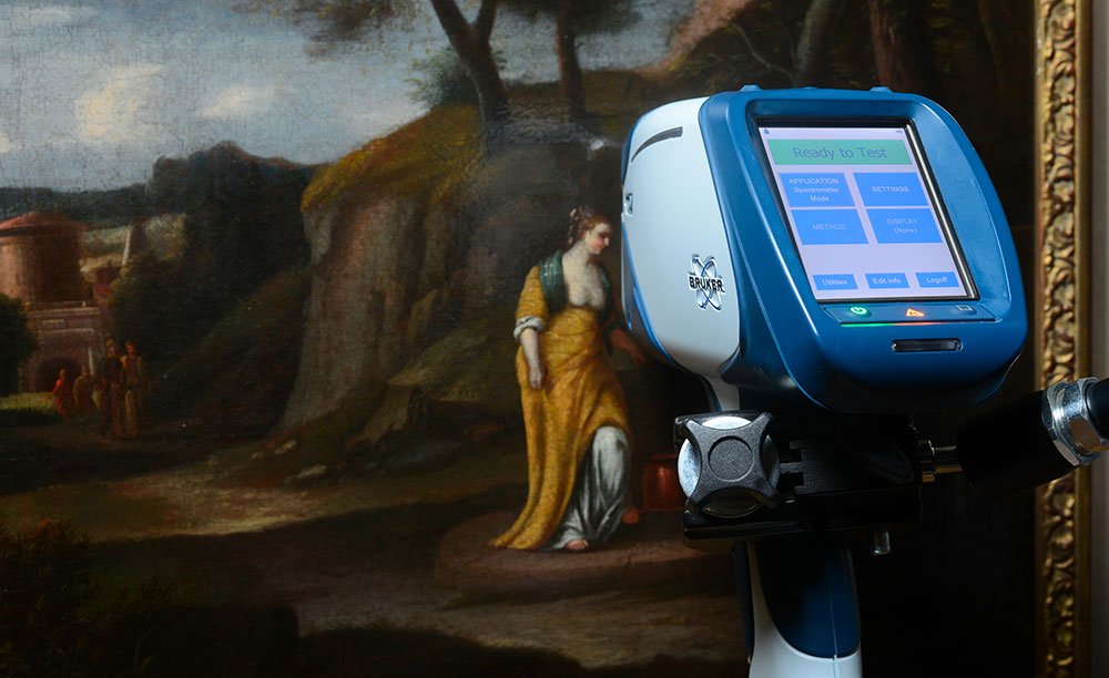
XRF IN SITU SPECTROSCOPY
The portable instrument lets us carry out XRF analysis in situ and obtain qualitative and semi-quantitative information on the elements present in the artefact.

RAMAN SPECTROSCOPY IN SITU
The portable instrument lets us carry out micro-Raman analysis in situ and obtain qualitative, molecular information on pigments and materials found in the artefact.

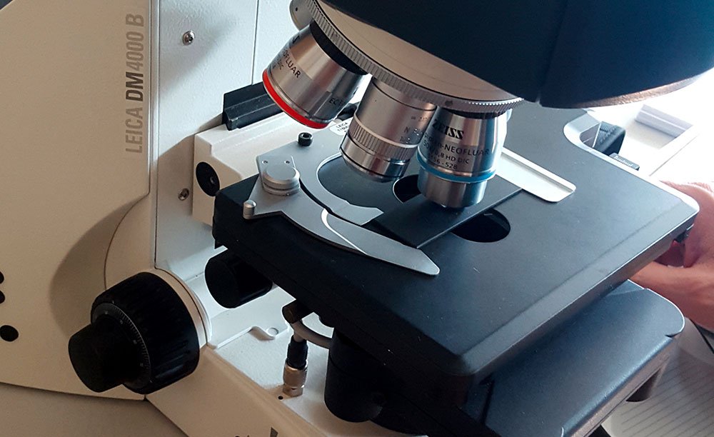
DIGITAL MICROSCOPY FOR THE RECOGNITION OF MATERIAL MORPHOLOGY AND 3D SURFACE RECONSTRUCTION
This lets us accomplish a microscopic and micrometric analysis of the surface of an artefact, by giving information on the morphological nature of the constituent materials.
INVASIVE
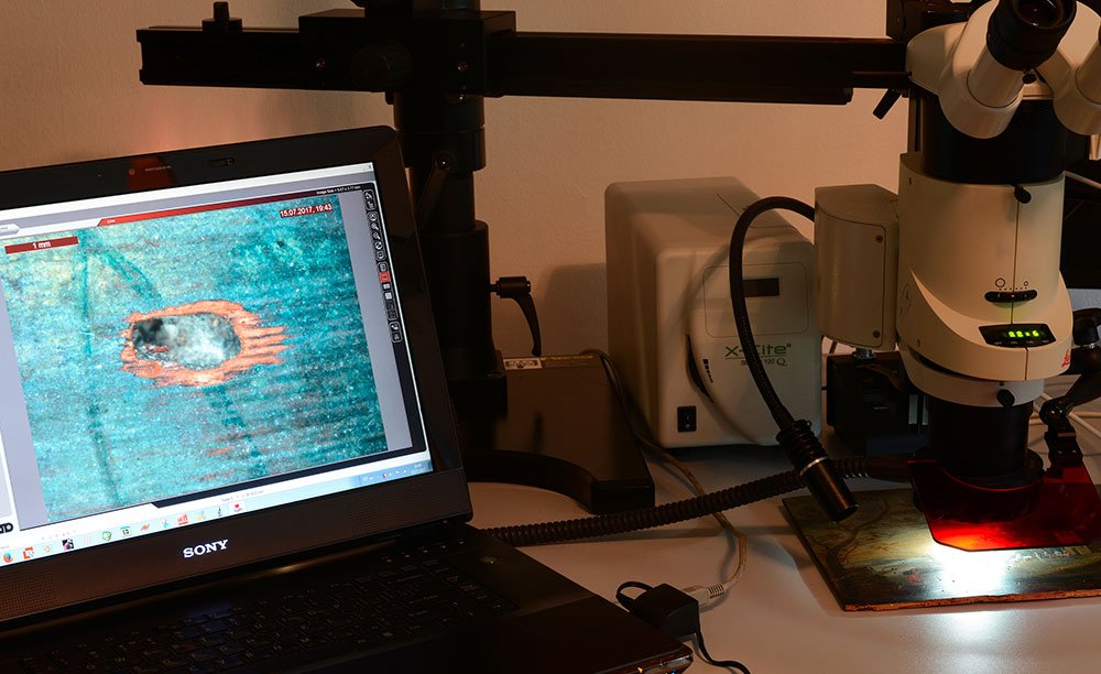
MICRO-SAMPLE EXTRACTION PROCESS
Through the use of a stereomicroscope, it is possible to extract micro-samples that are representative of both the surface and the deeper layers of a work of art.

TRAPPING MICRO-SAMPLES IN RESIN; CREATION OF THIN SECTIONS AND STRATIGRAPHIC CROSS-SECTIONS
This allows the creation of cross-sections that can be studied directly under the microscope to evaluate their stratigraphy or their mineralogical typology.
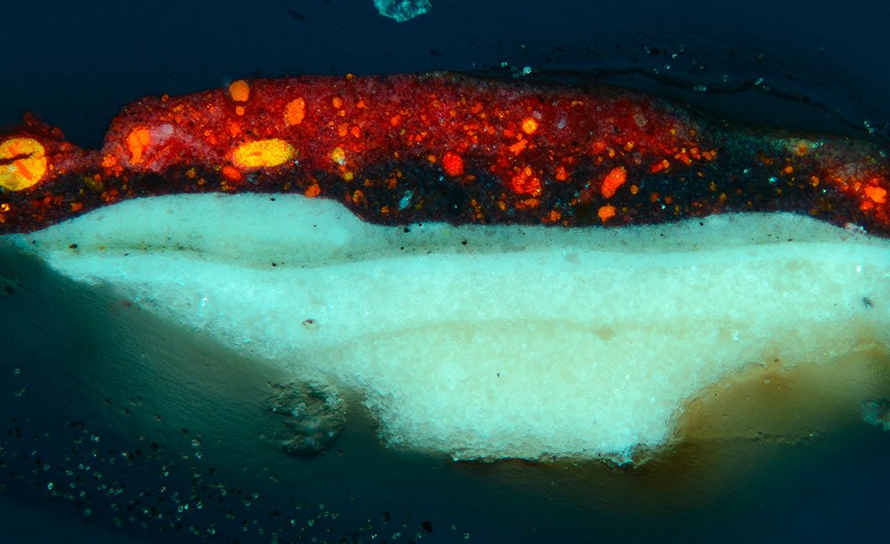
MULTISPECTRAL STUDY AND PHOTOGRAPHY OF MICROSCOPIC SECTIONS
Allows us to take images using visible, UV, infrared and false colour infrared to study the stratigraphy of a work of art.
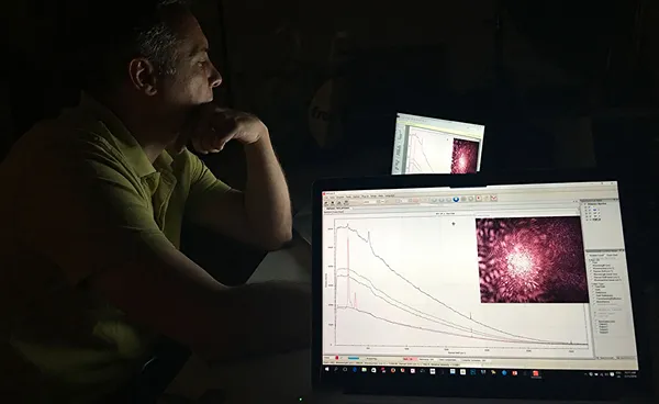
MICRO-RAMAN SPECTROSCOPY ON SAMPLE AND SECTION
This provides us information about the nature of the pigments and other materials of a work of art present in a cross-section.
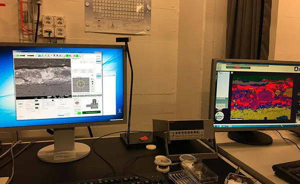
SEM-EDS SECTION ANALYSIS
This provides information on the nature and distribution of the elements of an artwork in the cross-section.

DATING USING C14 METHOD
The C14 method allows us to date organic materials such as wood, textile fibers, and paper.
INSTITUTIONS WE WORK WITH


